NEWS

Инсулиново лечение

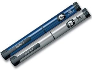
  • insulin pen picture

Какво представлява инсулиновата терапия?

Инсулинът е хормон, който панкреасът ви произвежда, за да позволи на клетките да използват глюкозата. Когато организмът Ви не произвежда или не използва инсулина както трябва, можете да приемате произведен инсулин, за да контролирате кръвната си захар. Повечето хора приемат инсулин, като го инжектират в кожата, въпреки че има и версия, която се вдишва. Ако имате диабет тип 1, се нуждаете от инсулин, защото панкреасът ви вече не произвежда хормона. Ако имате диабет тип 2, може да се нуждаете от инсулин, въпреки че много хора с тази форма на заболяването могат да контролират кръвната си захар без него.

insulin pen picture

Какво представлява инсулиновата терапия?

Инсулинът е хормон, който панкреасът ви произвежда, за да позволи на клетките да използват глюкозата. Когато организмът Ви не произвежда или не използва инсулина както трябва, можете да приемате произведен инсулин, за да контролирате кръвната си захар. Повечето хора приемат инсулин, като го инжектират в кожата, въпреки че има и версия, която се вдишва. Ако имате диабет тип 1, се нуждаете от инсулин, защото панкреасът ви вече не произвежда хормона. Ако имате диабет тип 2, може да се нуждаете от инсулин, въпреки че много хора с тази форма на заболяването могат да контролират кръвната си захар без него.

Какви са различните видове инсулин?

Много форми на инсулин лекуват диабет. Те се групират според това колко бързо започват да действат и колко дълго трае ефектът им. Видовете инсулин включват:

Бързодействащи:

Този вид инсулин започва да действа след около 15 до 20 минути и продължава от 1 до 5 часа, в зависимост от вида, който използвате. Приемате бързодействащ инсулин преди хранене и той обикновено се съчетава с по-дългодействаща форма на инсулин.

Дълготрайно действие:

Тази форма осигурява инсулиново покритие за цял ден. Вероятно ще използвате по-краткодействащ тип инсулин с нея.

Предварително смесен:

Съществуват няколко форми на предварително смесен инсулин, включително Humulin, Novolog и други. Тези разновидности комбинират краткодействащи и среднодействащи инсулини в една бутилка или инсулинова писалка, което някои хора намират за по-лесно за прилагане.

Кога се поставя инсулина?

Бързодействащи инсулини: Около 15 минути преди хранене
Късодействащи инсулини: 30 до 60 минути преди хранене
Среднодействащи инсулини: До 1 час преди хранене
Предварително смесени инсулини: В зависимост от продукта, между 10 минути или 30 до 45 минути преди хранене


Най-добрите места за инжектиране на инсулин

Мястото на тялото, където си поставяте инжекцията, може да е от значение. Инсулинът ще се усвои най-равномерно, когато го инжектирате в корема си. Следващите най-добри места за инжектиране са ръцете, бедрата и седалището. Създайте си навик да инжектирате инсулина в една и съща обща област на тялото си, но променяйте точното място на инжектиране. Някои лекари препоръчват местата за инжектиране да се разминават поне на ширината на един пръст или малко повече, ако използвате инсулинова помпа. Това помага да се намалят белезите под кожата. Ако на мястото на инжектиране се появят твърди бучки, може би имате белези и трябва да използвате друго място за инжектиране.


Какъв тип инсулин е най-подходящ за моя диабет?
Вашият лекар ще работи с Вас, за да Ви предпише типа инсулин, който е най-подходящ за Вас и Вашия диабет. Този избор зависи от много неща, включително:

Как реагирате на инсулина. Колко време е необходимо на организма да го абсорбира и колко дълго той остава активен, варира при различните хора.
Изборът на начин на живот. Видът на храната, която консумирате, количеството алкохол, което пиете, или количеството физически упражнения, които правите, ще повлияят на начина, по който тялото ви използва инсулина.
Вашето желание да си правите по няколко инжекции на ден
Колко често проверявате кръвната си захар
Вашата възраст
Вашите цели за управление на кръвната Ви захар
Вашият лекар може да Ви предпише повече от един вид. Може да се наложи да приемате инсулин повече от веднъж дневно, да разпределите дозите си през деня или да добавите други лекарства.


Странични ефекти на инсулина
Основните странични ефекти включват:

Ниска кръвна захар
Увеличаване на теглото, когато започнете да го използвате за първи път
Бучки или белези на местата, където сте си направили твърде много инжекции
Обрив на мястото на инжектиране или, рядко, по цялото Ви тяло

Заключение

Съществуват много форми на инсулин, които се различават по това колко време им е необходимо, за да започнат да действат, да достигнат максималната си ефективност и да спрат да действат. Вашият лекар може да ви помогне да определите кой инсулин или комбинация от различни видове инсулин е подходящ за вас. Съществуват и различни начини за приемане на инсулин, така че можете да изберете варианта, който ви е удобен и най-лесен за употреба.

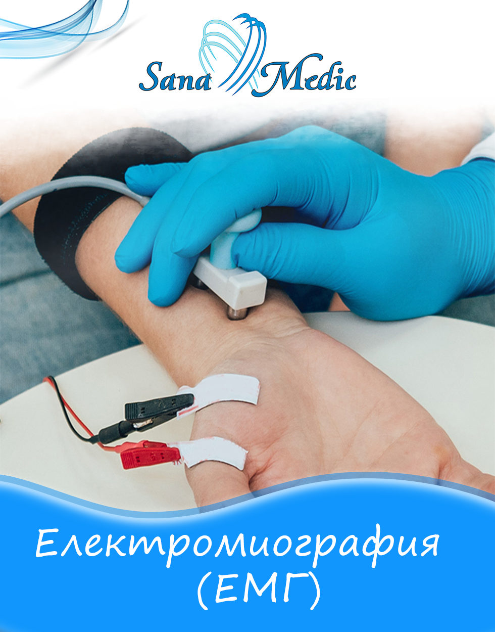
Electromyography (EMG)

neurology, неврология

Electromyography (EMG) is a diagnostic procedure to assess the condition of muscles and the nerve cells that control them (motor neurons). EMG results may reveal nerve dysfunction, muscle dysfunction, or problems with signal transmission from nerve to muscle.


Motor neurons transmit electrical signals that cause muscles to contract. In an EMG, tiny devices called electrodes are used to convert these signals into graphs, sounds or digital values, which are then interpreted by a specialist.

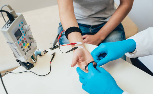
During a needle EMG, an electrode inserted directly into a muscle records the electrical activity in that muscle.

Nerve conduction studies, another part of EMG, use electrode stickers placed on the skin (surface electrodes) to measure the speed and strength of signals traveling between two or more points.

For more information about the team of neurologists at SanaMedic Medical Center, visit the services section of our website!

How do you prepare?


Food and medicine
When scheduling an EMG, ask if you should stop taking prescription or over-the-counter medications before the test. If you are taking a medicine called Mestinon (pyridostigmine), you should specifically ask if this medicine should be stopped for the study.

Bathing
Take a shower or bath shortly before the examination to remove oils from your skin. Do not apply lotions or creams before the examination.

Other precautions
The nervous system specialist (neurologist) who performs the EMG will need to know if you have certain diseases. Tell the neurologist and other EMG lab staff if:

  1. you have a pacemaker or other electrical medical device
  2. taking blood thinners
  3. Have haemophilia - a bleeding disorder that causes prolonged bleeding

Before the procedure
You will probably be asked to change into a hospital gown for the procedure and lie on the examination table. To prepare for the examination, the neurologist or technician will place surface electrodes in different locations on your skin depending on where you are experiencing symptoms. Or the neurologist may place needle electrodes in different places depending on your symptoms.

ЕМГ, неврология, emg, neurological examination

During the procedureа

When the study is in progress, the surface electrodes will sometimes transmit a small electric current that you may feel as a prickling or spasm. The needled electrode may cause discomfort or pain, which usually subsides soon after the needle is removed.

During the needle EMG, the neurologist will assess whether there is spontaneous electrical activity when the muscle is at rest - activity that is not present in healthy muscle tissue - and the degree of activity when you gently contract the muscle.

He or she will give you instructions for resting and shortening the muscle at appropriate times. Depending on what muscles and nerves the neurologist is examining, he or she may ask you to change your position during the examination.

If you are concerned about discomfort or pain at any time during the study, you may want to talk to the neurologist about taking a short break.

After the procedure


You may have temporary, minor bruising where the electrode was inserted into the muscle. These bruises should disappear within a few days. If they continue to appear, contact your GP.

Results


The neurologist will interpret the results of your examination and prepare a report. Your primary care physician or the physician who ordered the EMG will discuss the report with you at a follow-up appointment.

Изотчници:

  1. Rubin DI. Needle electromyography: Basic concepts. Handb Clin Neurol. 2019;160:243-256. doi: 10.1016/B978-0-444-64032-1.00016-3. PMID: 31277852.
  2. Dondelinger RM. Electromyography–an overview. Biomed Instrum Technol. 2010 Mar-Apr;44(2):128-31. doi: 10.2345/0899-8205-44.2.128. PMID: 20586391.

Intermittent fasting

  • сънна апнеа

Tables and tempting dishes are a common companion of Christmas holidays not only in Bulgaria. After the Christmas season usually comes the time of New Year promises for strict diets. The usual uncertainty about what approach to take for weight reduction. There is an endless amount of information about different diets, both on the internet and in self-improvement books, advice from fitness instructors and even various recommendations for weight-reducing medication.


fasting, time, intermittent fasting, minimalist


One of these approaches is intermittent fasting. This is an approach where calorie intake is restricted, but the focus falls on the amount of time a person can consume food, within a day or week.
One option is to alternate a day with food intake with one during which you fast. The other option, more commonly used, is to alternate 16 hours of fasting with 8 hours of meal time in a 24-hour period. Although both calorie restriction and intermittent fasting can lead to an overall reduction in caloric intake, this is not the focus of intermittent fasting.

As is evident from the name of the approach - intermittent fasting - it is not only the eating period that is important, but also the "fasting". The word in this case is somewhat misleading, as it is not the feeling of hunger that is significant, but the lack of calorie intake during this period. During the fasting period (the 16 hours) the intake of unsweetened teas, water, coffee without sugar is allowed.

How does intermittent fasting work?

The main hypotheses for the effect of fasting on the tissues of the human body are three: 1) reduction of oxidative stress; 2) synchronization of caloric intake with circadian rhythms; 3) ketosis. [1]

The oxidative stress hypothesis is supported by studies that have shown fewer free radicals produced by mitochondria after caloric restriction [2].

Circadian rhythms are changes in biological processes (e.g. hormone secretion) within a 24-hour period. An example is that insulin levels are higher in the morning than in the evening. In view of this, it is suggested that food intake between 8:00 and 16:00 is more favourable compared to eating only in the afternoon hours. Such an approach has been shown to be balgosprious in reducing blood pressure and weight [3].

Analogous to the ketogenic diet, intermittent fasting also increases ketone production. This is a sign that the metabolism begins to use fatty acids, therefore breaking down adipose tissue.

What are the benefits of intermittent fasting?

Intermittent fasting has positive effects on multiple cardiovascular risk factors, including obesity, hypertension, dyslipidemia, and diabetes. In addition, there is evidence of a better prognosis in patients who have already had a heart attack.

In SanaMedic there are specialists who can help you with a diet plan!

Източници:

1. Dong TA, Sandesara PB, Dhindsa DS, Mehta A, Arneson LC, Dollar AL, Taub PR, Sperling LS. Intermittent Fasting: A Heart Healthy Dietary Pattern? Am J Med. 2020 Aug;133(8):901-907. doi: 10.1016/j.amjmed.2020.03.030. Epub 2020 Apr 21. PMID: 32330491; PMCID: PMC7415631.
2. Merry BJ. Oxidative stress and mitochondrial function with aging–the effects of calorie restriction. Aging Cell. 2004 Feb;3(1):7-12. doi: 10.1046/j.1474-9728.2003.00074.x. PMID: 14965349.
3. Moro T, Tinsley G, Bianco A, Marcolin G, Pacelli QF, Battaglia G, Palma A, Gentil P, Neri M, Paoli A. Effects of eight weeks of time-restricted feeding (16/8) on basal metabolism, maximal strength, body composition, inflammation, and cardiovascular risk factors in resistance-trained males. J Transl Med. 2016 Oct 13;14(1):290. doi: 10.1186/s12967-016-1044-0. PMID: 27737674; PMCID: PMC5064803.

Doppler examination

  • доплер изследване варна

What is a Doppler study ?

доплер изследване варна

Doppler sonography is a harmless and painless method for examining the movement of blood in the blood vessels- arteries and veins. Its basis is the study of the frequency of ultrasound waves. There are three zones of Doppler examination -Transcranial, Extracranial and Peripheral Doppler Sonography.

Types of Doppler examination

Extracranial Doppler sonography - examines the blood vessels and blood flow in the cervical region and skull (head Doppler).

Peripheral sonography is a diagnostic procedure that examines veins and arteries of the extremities - Doppler can be performed on the legs and also on the arms.

Transcranial sonography - ultrasound beams penetrate the skull and practically reach all blood vessels in the brain. This makes it possible to directly track blood flow in the intracranial cerebral arteries and obtain a comprehensive picture of cerebral blood flow, vascular resistance, the amount of blood that passes per unit time, blood vessel constrictions, the presence of atherosclerotic plaques and thrombi. Investigated is the movement of blood in the vessels, the qualities of its flow, direction and speed and determining the state of the vascular bed, the patency of the vessels, abnormalities in the walls and along the course of the various arteries and veins, including the imaging of atherosclerotic plaques.

доплер сонография варна

доплер изследване варна

For the purpose of neurology, Doppler examination is associated with extracranial and transcranial areas and is indicative mainly in:

Conditions:

– - Headaches (migraines);

– - spasms of blood vessels;

– - problems with dehydration of the limbs and numbness;

– - diabetic polyneuropathy;

– - dizziness and balance disorder;

– - stroke /hemorrhagic or ischemic/;

– - congenital anomalies of the cerebral arteries;

– chronic peripheral arterial circulatory insufficiency;

- varicose veins

– - thrombophlebitis, etc.

Advantages of the method:

– The examination is performed using ultrasound, making it safe and without any pain for the patient

– Through Doppler sonography, blood vessels can be examined in their entirety - this includes accumulated plaques, dilations and any interference that prevents proper irrigation and blood supply.

– The method allows tracking the type and degree of disability and making predictions about its development

Contraindications:

The test has no practically established contraindications, which makes it a much preferred, efficient and widely applicable diagnostic method that is safe for the patient.

доплер сонография варна

Doppler examination in SanaMedic Varna ?

доплер изследване варна

The duration of the examination varies - from a few minutes to just over half an hour - depending on the number of vessels examined and also the area.

MC SanaMedic has a state-of-the-art ultrasound scanner, which enables a perfectly precise diagnosis of all changes in the blood vessels connected to the brain.

The capabilities of the specialists working at SanaMedic Varna are also related to the study of the peripheral cardiovascular network. This type of examination-comprehensive allows a comprehensive diagnosis of existing and possible cardiac and vascular problems.

More detailed information about the state-of-the-art ultrasound scanner we have in our centre can be found in the following link:

сонография варна


Медицински център ВарнаDoppler Examination Varna

Sleep apnoea

  • сънна апнеа

What is sleep apnoea?

сънна апнеа

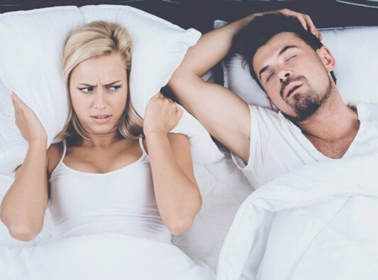
Sleep apnea (apnea - from Latin for lack of breathing, cessation of breathing) is a common condition characterized by episodes of cessation of breathing during sleep. Breathing stoppages can last from a few seconds to minutes and occur 5 to 30 or more times per hour. After a time , normal breathing returns, and may be accompanied by loud snoring or choking sounds.

Sleep apnea is one of the commonly seen chronic diseases that disturbs sleep 4 or more times a week.

This leads to poor sleep quality and fatigue during the day. Sleep apnea is one of the main causes of sleep deprivation, lack of quality sleep and daytime tiredness and fatigue.

The problem in diagnosing the disease is that it only manifests itself at night, which is why the person often does not know about it. A family member and/or bed partner may notice its first signs.

Sleep apnea often cannot be diagnosed in a routine medical examination. For this purpose, there are special devices that record heart, breathing and brain activity during sleep. The information they accumulate is then relied upon by a specialist who should recommend a further course of treatment.

What are the risk factors andcauses for  sleep apnoea?

Risk factors for obstructive sleep apnea include:

–-male gender

–-high blood pressure

–-obesity

–-smoking

–-old age

–-use of narcotics, sleeping pills or alcohol

–-neurological diseases

–DIabetes

What are the symptoms ?

Symptoms of obstructive sleep apnea include:

–-heavy snoring

–-waking accompanied by gasping

–-morning headache

–Insomnia

–daytime sleepiness

–-concentration disorders

–-high blood pressure difficult to control

сънна апнея

How is sleep apnea diagnosed?

Previously, the main way to diagnose sleep apnea was so-called Polysomnography, which was only performed in the laboratory. In this examination, you sleep overnight in the laboratory, during which time machines are attached to you that monitor organ function, breathing patterns, and leg and arm movements. Advances in medical technology now allow sleep apnea diagnosis to be done entirely at home, without having to leave the comfort and convenience of your home. This is possible with the help of modern portable sleep and breathing disorder screening devices. These devices perform complete respiratory monitoring of vital parameters that indicate the presence of sleep disturbances without having to change your daily routine.


Treatment methods for obstructive sleep apnea?


Промени в начина на живот:

сънна апнеа

–smoking cessation

–stopping alcohol use or not drinking alcoholic beverages a few hours before sleep

–losing weight

–avoid sleeping on your back

Sleep apnea machine treatment:

сънна апнеа

-CPAP mask (Continuous Positive Airway Pressure). This is the most widely used method of treating sleep apnoea (obstructive or central) and involves wearing a mask around the nose during sleep. The mask is connected to a machine that generates pressurized air, thus preventing the airway from closing.

-BPAP mask (Bilevel Positive Airway Pressure - Positive Airway Pressure at two levels) - unlike CPAP, which maintains a constant supply air pressure when you inhale and exhale, BPAP machines send air at a higher pressure when you inhale and at a lower pressure when you exhale. The purpose of this type of treatment is to help weak breathing. Some BPAP machines automatically supply air when they sense that you have not breathed over a certain number of seconds.

oxygen therapy - The machine supplies extra oxygen to your respiratory system while you sleep. This therapy has shown effectiveness in some cases of sleep apnea.

-rTMS (repetitive transcranial magnetic stimulation) Transcranial magnetic stimulation is a new non-invasive method of modulating the activity of the central nervous system. The idea of the method is to achieve an overall improvement of the condition without the use of drugs, but entirely naturally. It is used for insomnia, burnout syndromes, depression, memory and concentration disorders, after strokes and other brain damage.

Сънна апнея

What complications can sleep apnea cause?

Untreated sleep apnea increases the risk for:

–The development of high blood pressure, heart attack, stroke, obesity and diabetes;

–The development or worsening of heart failure;

–The development of cardiac arrhythmias;

–Fatigue - due to frequent awakenings at night and can lead to reduced concentration at work or while driving, which can lead to serious accidents

Clinic of Sleep Medicine and Sleep Apnea at SanaMedic Medical Center Varna

The office has state-of-the-art equipment - electro-encephalograph (EEG), electrocardiograph (ECG), electromyograph (EMG), transcranial magnetic modulator (rTMS). сънна апнея

SanaMedic and Miss Varna 2020

мис варна 2020

On 26 and 27 August this year was held one of the most prestigious beauty and style events in our maritime capital - the Miss Varna 2020 Contest. MC SanaMedic had the exclusive pleasure to be an official sponsor and jury member in this year's event.

As a center dedicated to the mission of caring for the beauty and health of every person, we had the responsible task of helping the contenders in their battle for the crown. One of our dermatologists, Dr. Gloria Dimitrova, gave the girls valuable tips on what are the most common mistakes we make when we take corrective action on ourselves; what are the important habits that keep our skin youthful and taut for long. She also talked about the risks that accompany some of the most common aesthetic enhancements, as well as ways to avoid them. See how the beauty ambassadors spent their time at our medical center and how, we were able to help them become their best selves:

And here is a short interview with dermatologist Dr. Gloria Dimitrova as one of the jury members of Miss Varna 2020.

Медицински Център Варна

Nodular goiter

The article was written by Assoc. Prof. M. Siderova and Prof. K. Hristozov
д-р Мира Сидерова

Nodular goiter is a heterogeneous disease in clinical, physiological and histological aspects: nodules can be single or multiple, accompanied by hyper- or hypofunction, benign or malignant. The main tasks of the clinician in the detection of nodular goiter are the exclusion of malignancy and of hyperthyroidism due to thyroid autonomy.

Epidemiology

Thyroid nodules are detected in approximately 5% of the population using palpation as a screening method. When ultrasound screening is performed, this percentage increases tenfold and reaches between 20 and 75% of the general population. A higher incidence of thyroid nodules is seen in areas endemic for goiter. Thyroid nodules are rare in children and adolescents, and their incidence increases linearly with age. Women are affected 2-4 times more than men.

Thyroid carcinoma is a rare human malignancy, accounting for up to 1% of malignancies, but is the most common endocrine cancer. It is found in about 5% of thyroid nodules. Over the last 30 years, the annual incidence of thyroid carcinoma has increased from 6 to 8.7/100,000, i.e. 2.4 times. This increase is thought to be largely due to improved diagnosis and the increase has been at the expense of diagnosing nodules less than 2 cm, with the mortality rate from thyroid carcinoma unchanged at 0.5/100,000. The accident in
Chernobyl in 1986 marked a new era in the incidence of thyroid cancer. The risk of developing thyroid cancer, especially in Ukraine, Belarus, western Russia and neighbouring countries, was greatest for children who were then under 9 years of age and especially under 5 years of age and had probably ingested a large dose of radioactive iodine through milk and dairy products. An increasing number of thyroid carcinoma cases are expected in Japan in the coming years following the Fukushima accident.

Clinical Evaluation

From the history, the most important information is the sex (men- they are at higher risk for malignant lesions), as well as the age of onset of the nodule. Risk age groups are those under 20 years and over 70 years. Past radiotherapy to the head and neck is also a risk factor. Dysphonia, dysphagia and dyspnoea as well as rapid growth of the nodule are alarming symptoms. Familiality should also be sought, especially in relatives with medullary, papillary carcinoma u MEN type 2 (multiple endocrine neoplasia). Symptoms of hyperthyroidism as well as use of medications containing iodine may suggest toxic nodular goiter. Most nodules are asymptomatic and the absence of symptoms does not rule out malignancy.

Physical examination of the neck is mandatory, focused on the size, localization, consistency of the thyroid nodule, fixation to the surrounding tissues, the presence of neck pain, as well as enlarged cervical lymph nodes (LNs).

Laboratory examination

Most patients with benign or malignant thyroid nodules are euthyroid. However, serum TSH testing is appointed in all patients with a thyroid nodule. If the TSH is low, laboratory tests are expanded to include FT4 and FT3, which clarify whether it is overt (with elevated peripheral hormones) or subclinical hyperthyroidism (normal peripheral hormones combined with low TSH). In both cases, scintigraphy is appropriate to determine functional nodal activity. Most guidelines do not recommend scintigraphic hot node biopsies because they are rarely malignant. In high TSH, testing for FT4, anti-TPO, and anti-TG antibodies is indicated because autoimmune thyroiditis is the most common cause of hypothyroidism.

The serum thyroglobulin test has no place in the diagnosis of nodular goiter, since it is elevated in any gland enlargement, as well as in destructive changes. Its use as a tumor marker in patients treated for thyroid carcinoma makes sense only after a total thyroidectomyl has been performed.

The serum calcitonin assay is a marker for medullary carcinoma and correlates well with disease prevalence. Its routine measurement in the presence of a thyroid nodule is still controversial, as medullary carcinoma is found in less than 0.5% of nodules, and serum calcitonin is often false-positive in renal insufficiency or use of proton inhibitors. Its investigation is due in familial or clinical suspicion of medullary carcinoma or MEN type 2. If calcitonin is elevated, it is first repeated, followed by a stimulation test with pentagastrin or calcium.


Ultrasound diagnostics

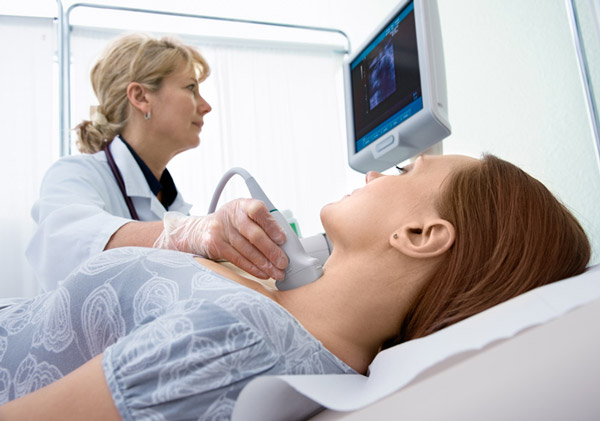
Ultrasound (US) examination is the most sensitive method for the detection of thyroid nodules, to accurately determine their size and structure. Ultrasonography is mandatory in patients with clinically established risk of thyroid carcinoma, in cervical lymphadenomegaly and in any palpable nodule. In 20% to 48% of patients with a single palpable nodule, other nodules are detected sonographically, i.e., polynodous stroma. The risk of carcinoma is the same in patients with a solitary nodule and a multinodular goiter. Moreover, carcinoma may lie in the dominant node (the one with the largest size) as well as in the nondominant ones.

The focus of the US examination in nodular goiter is to look for features at risk for malignancy - hypoechogenic solid nodule structure, uneven contours, lack of halo, anterior-posterior greater than transversal size, microcalcifications, intravascular chaotic Doppler signals, cervical lymphadenomegaly.

Elastography is another application of U3 to study the deformation and elasticsp of tissues in compression. The nodes or parts of nodes that deform minimally, i.e. are rigid and inelastic, are at risk.

There are also some ultrasonographic features suggestive of a benign nature of the nodule - clear borders, the presence of gentle continuous halo, dorsal acoustic enhancement, hyper- or anechogenic nodule structure, and the presence of eggshell-type macrocalcifications. Very often the pseudonodous form of Hashimoto's autoimmune thyroiditis is confused with polynodous goiter .

Fine-needle aspiration biopsy (FNA) is the most accurate and important diagnostic method to differentiate benign from malignant thyroid nodules. Routine performance of FNA has changed the management of thyroid nodules by avoiding unnecessary surgery of benign lesions and thus reducing the cost of treatment. On the other hand, preoperative FNA and demonstration of thyroid carcinoma implies a single-stage operation, total thyroidectomy, in contrast to multiple operations in patients without preoperative cytological clarification. The sensitivity of FNA ranges between 65% and 98% and the specificity between 72% and 100%. There are different methods of performing FNA - with and without aspiration, "free hand", and FNA under ultrasonographic control, the latter having advantages in nonpalpable nodules as well as in the choice of the biopsy site from solid-cystic nodulesl.

Cytology results from TAB are classified into five diagnostic categories:

Class 1. Nondiagnostic

Reasons for insufficient cytological material can be very small nodules, poor in epithelial cells and erythrocyte-rich sample, poor fixation, sclerotic lesions (Hashimoto's thyroiditis-fibrosing variant), cystic degeneration of the node, necrosis. In case of one non-diagnostic result of TAB, it should be repeated. In case of a second non-diagnostic TAB of a solid nodule, most authors recommend surgical removal of the nodule. This decision is also influenced by the ultrasonographic characteristics, size and dynamics in nodule growth.

Class 2. Benign

In case of a benign cytological result, the patient is followed clinically, c TSH u US at 6 to 18 months. Repeat FNA is recommended when clinical or US features suggestive of malignancy occur or when nodule volume increases more than 50% from baseline. Suppressive levothyroxine treatment of benign nodules used in the past is not routinely recommended because of its low efficacy and nonbeneficial cardiovascular and bone effects. Indications for surgical treatment of benign thyroid nodules are compression syndrome, previous external irradiation, rapid growth, susceptible U3 features, and cosmetic considerations. The extent of resection for a single nodule is lobectomy with ischemectomy, and for a multinodular goiter is near-total thyroidectomy. Alternatives to surgical treatment are percutaneous ethanol sclerosant, laser ablation, and high-frequency ultrasound (H1FU) ablation. Radioiodine therapy (RIA) is the treatment of choice for hyperfunctioning and/or symptomatic nodules in patients with high operative risk. Follow-up of patients after RYL for stromal nodosis includes thyroid function testing (TSH, FT3, FT4) and U3 every 3-6 months, and RIA can be repeated in cases of persistent hyperthyroidism or insufficient volume reduction in compressive manifestations.

Class 3. Folicular nodules

Repeat biopsy is not recommended for this category of nodules, as cytological examination cannot differentiate follicular adenoma from follicular carcinoma (respectively Hurthle cell adenoma from carcinoma). Differentiation requires histological verification of capsular and/or vascular invasion. Several molecular markers (Galectin-3, Cytokeratin-19, Fibronectin, Claudin, HBME1) have been identified that have different expression in benign and malignant thyroid nodules. Their immunocytochemical study in FNA material could increase the sensitivity and specificity of routine cytological examination. Most guidelines recommend operative removal of all follicular lesions.

Class 4. Suspicious

Patients with diagnostic category 4-specific thyroid nodules are referred for surgical treatment. For these nodules, intraoperative geographic examination is also recommended.

Class 5. Malignant

Surgical removal is mandatory in case of a malignant cytological result. Preoperatively, a good US view or CT scan for cervical lymph nodes is necessary. In the case of a suspected metastatic LNs, thyroglobulin or calcitonin in the smear from the needle used to perform the FNA of the LNs can be tested along with its cytology. CT/MRI is indicated in selected cases to determine invasion of the trachea and surrounding structures.

See section "Endocrinology" (link)

logo Sana Medic Varna

CONTACT US FOR PROFESSIONAL CONSULTATION AND TREATMENT.

tel. 052/608 313
mobile: 0877 868 864

Adress: Varna, "Sofia" No. 5 street

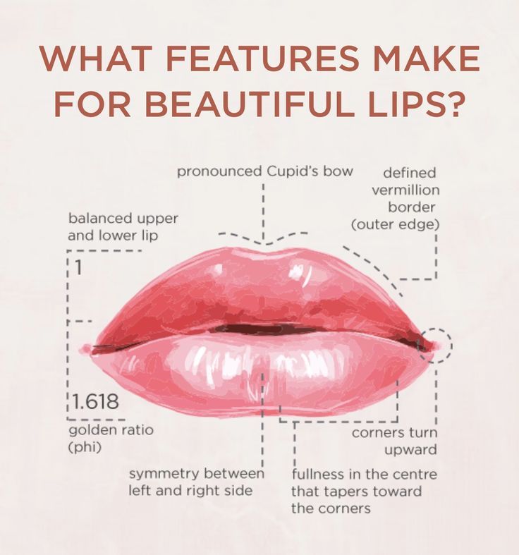
Hyaluron in the lips - what you need to know

*The article was edited by dermatologist Dr. Gloria Dimitrova

хиалурон в устните

Before you put hyaluron in your lips!

Plump cheekbones, voluminous lips, smoothed skin - some get them by birth (we can't blame them), others with a little help from the dermatologist and a hyaluron in the lips or some other place on the face. And if watching your favorite TV host or live from a famous influencer you can't guess which of the 2 categories they fall into- well, that's exactly the idea of a properly placed hyaluronic filter.

Placement of hyaluron filter is the most used method for volumizing and shaping various parts of the face in recent years (especially hyaluron in the lips), mainly due to the fact that the procedure is quick and the effects are instant. The method is also one of the safest- here we are proceeding on the premise that you have done your research and are leaving yourself in the hands of an educated and proven professional.

In this regard, to help you in the research part, we have prepared this article for you with information on the things you should know before proceeding to the hyaluron placement in the lips or any other place on your face.

Difference between hyaluron and botox?

To begin with, we would like to make two basic distinctions that sometimes cause patient confusion and clarify the logic behind each of them - the difference between hyaluron and botox.

Botox is a toxin that prevents the muscle from contracting-"paralyzes" it-and thus prevents it from forming wrinkles and lines.

The action of Hyaluronic acid is quite different. Its role is to fill the lost volume. With the aging process, the amount of natural hyaluronic acid in the body becomes less and as a result, the skin roughens and loses elasticity. By injecting hyaluronic acid, the lost volume is restored and a tightening effect is achieved, which clears lines and wrinkles. The placement of hyaluron in the lips, on the other hand, aims to give more volume.

See more about the two procedures

хиалурон                                            хиалурон в устните

Main types of hyaluronic fillers

Now that we've clarified the difference, we'll tell you a bit about the basic types of hyaluronic fillers:

permanent - хиалурон в устните  they are composed of substances that are non-degradable for the body (most often silicone), which makes them permanent. This also makes them more dangerous- they have more side effects and carry a risk of malignancy. They are almost no longer used in aesthetic dermatology.

collagen stimulators- the difference from hyaluronic is that in addition to filling wrinkles on the skin, they aim to stimulate the synthesis of natural collagen. Stronger connective tissue is formed and the skin becomes plumper and firmer. The result is longer lasting.

Temporary the most used are those based on hyaluronic acid. It is an essential component of many organs in the human body, including our skin. Its main role is to be a natural hydrator- it maintains the volume of the skin thanks to its ability to attract and retain water within itself. Over time, the aging process causes its quantity to decrease, its volume to drop and wrinkles to appear. Hyaluronic acid injection aims to restore the lost hyaluronic acid. And because it is a natural part of our body, over time it is absorbed by the body without causing any unwanted side effects.

Why is putting hyaluron in the lips the preferred method?

The main reason this is the most preferred method is that it allows adding volume or removing the appearance of wrinkles in just a few minutes spent at the doctor. Another great advantage is the great leeway they provide the dermatologist- in case your lip volume is not enough, it is not a problem to add extra hyaluron, or if there are asymmetries, they can be easily corrected. In the reverse hypothesis, if the volume obtained is more than desired or the final result is unsatisfactory- hyaluronic acid can be easily broken down with the help of a special enzyme and thus removed.

хиалурон в устните

In turn, hyaluronic fillers (fillers) are divided into different groups. Depending on the type of effect that is intended; the area where it is applied and the depth of injection- there are differences in composition and structure. For example, if we need slight corrections for some fine lines we would use a filler with a lower density, the opposite is true when we are working on a more extensive area and need more visible volume- for example in the cheekbones.

The most important things to research before getting hyaluron in your lips!

Okay, now that we're clear on what the different types of fillers are and how they work it's time to focus on the next important aspect in your research - these are the brand of the fillers and the specialist who puts them on you. Among the most well-known brands , you have probably heard of are Juvederm; Restylane is another well-established brand. We at SanaMedic rely on the symbol of uncompromising quality and continuous innovation in the beauty industry - the Swiss-based Teoxane and their products  Teosyal. But whichever brand you decide to trust, the most important thing you should pay attention to is whether the product you are injecting is EU approved and has a safety certificate. This ensures that it has been produced using patented technology, has been carefully researched and tested for many years before being released on the market. This is something you should not compromise on as it has a direct bearing on your health and its safety.хиалурон в устните варна

As safe as a procedure is, remember that it is as safe as competent as the one who performs it. The insertion of hyaluron into the lips should be performed only by a qualified specialist-dermatologist, with sufficient experience and which is certified to operate with the products it puts you. A prepared doctor knows the anatomy of the human face and the location of the various blood vessels on it, he also knows how to react if necessary to prevent complications. Imagine the consequences if the procedure is performed by someone who does not know all the specifics and risks. Inserting hyaluron in places not designated for this purpose or even worse by persons without the necessary expertise expose your health to serious danger. After all, beauty is all about health, and health comes first.

Price for putting hyaluron in the lips?

We won't lie to you, hyaluron injections are not among the cheapest aesthetic adjustments you can get. If someone is trying to convince you otherwise and offer you prices that are too low- then is too good to be true. Prices vary according to different factors, but the basic rule is that the price of the procedure should not be below 400 BGN. - this is the minimum price to be able to count on some quality. Really good quality hyaluron is now in the range of 450 BGN and up.

Does it hurt and how long does the effect last?

Inserting hyaluron into the lips is not considered a painful procedure, in addition, some fillers also contain the ingredient lidocaine, which numbs sensitivity when injected. Also, the area is pre-anesthetized by applying anesthetic in the form of a cream.

The duration of the effect varies between 4 months to not more than 1 year4 months to not more than 1 year- depending on how you take care of yourself and how quickly your body will resorb the hyaluron.

хиалурон в устните

It is also important to know in which cases hyaluron should not be placed in the lips

Важно е също така да знаете в кои случаи хиалуронът не трябва да се поставя в устните.

-the presence of herpes and infections in the treated area. This increases the risk of spread when injected. In such cases, wait for these to subside before proceeding with insertion. Any ethical and experienced professional would refuse to inject you with hyaluron in such a condition.

- in certain immune diseases. Therefore, the procedure is always preceded by a consultation with a specialist.

- when pregnant or breastfeeding

These are the main factors to consider when making your choice. Our personal advice is to have "done your homework" and spend a long time in research and checking. Putting it in takes minutes, but the result will stay with you for months.

That is why the philosophy of SanaMedic has always been "Your health and beauty the focus of our efforts". Because we believe that safety comes first.

хиалурон

CONTACT US FOR PROFESSIONAL CONSULTATION AND TREATMENT.

tel. 052/608 313
mobile: 0877 868 864

Adress: Varna, "Sofia" No. 5 street

Репетативна Транскраниална Магнитна Стимулация

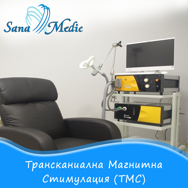
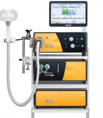
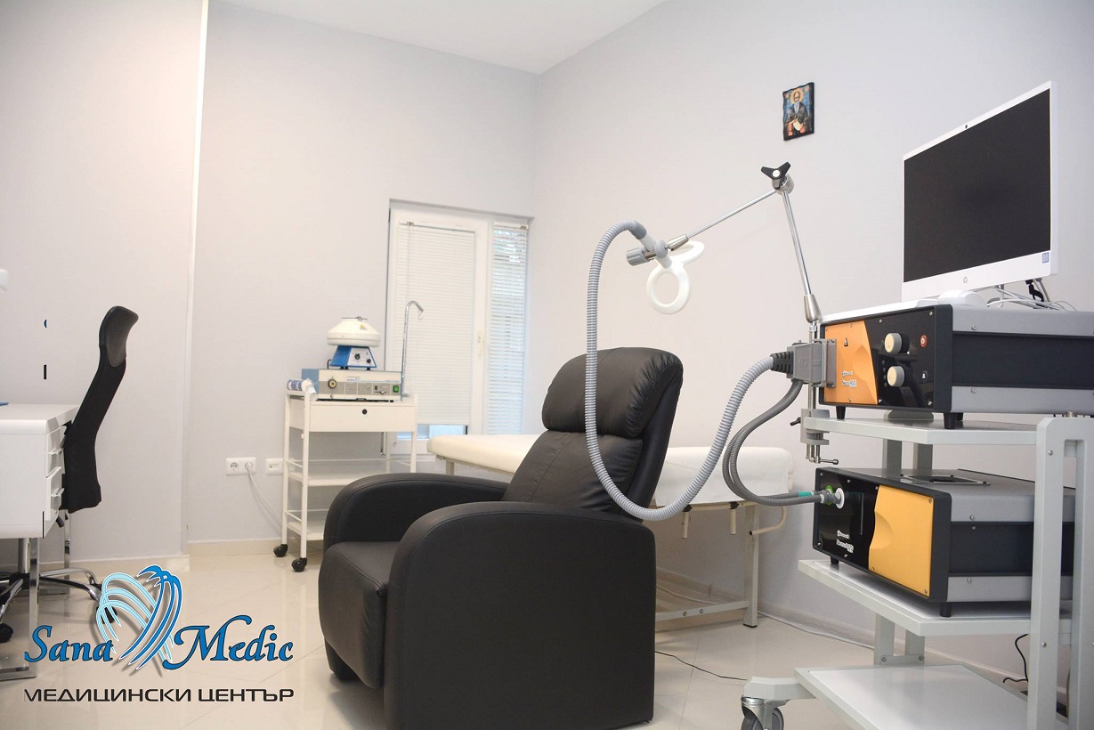
Transcranial magnetic stimulation (TMS) is a new non-invasive treatment method in which repetitive magnetic pulses are directed to specific areas of the brain. The idea of the method is to use a directed magnetic field in the activation or deactivation of specific areas of the brain. These magnetic pulses pass painlessly through the human skull and reach the brain cells, stimulating them and improving communication between different parts of the brain. When these pulses are repeated at regular intervals - this is known as Repetitive Transcranial Magnetic Stimulation (rTMS). The pulses are produced by placing a magnetic head in the area above the scalp - they are the same as the pulses produced by Nuclear Magnetic Resonance.

Conducting magnetic pulses into the brain probably sounds quite frightening to many of us, and probably also brings up associations from movies with startling shock therapy scenes that look more like torture than real treatment. ( The scene in question is from the movie One Flew Over the Cuckoo's Nest (1975) with Jack Nicholson). BUT we have taken the time for this article to convince you that ТМS has nothing to do with such type of conceptions - it is safe, painless and above all non-invasive method - ie no anesthetics, penetration into the body or recovery time. As a matter of fact, in a large percentage of cases the side effects are significantly less than those when using antidepressants.

Transcranial magnetic stimulation is a good alternative for patients in whom drug treatments are contraindicated or inappropriate because of their side effects, allergies, intolerance or because of pharmacophobia.


In what conditions is pTMS applicable?

Transcranial Magnetic Stimulation is a relatively new, non-invasive method used in the treatment of depression and other mental disorders.

High effectiveness is observed in patients with the following diagnoses:

  • Psychiatry:

☑ Treatment of depression;
☑ Anxiety disorders (Obsessive-compulsive disorder, Generalized anxiety disorder, Anxiety-depressive disorder, Stress reactions, etc.);
☑Increased anxiety and insomnia;
☑ Burnout syndrome (occupational exhaustion);
☑  Autism.

  • Neurology:

☑ Ischaemic strokes: appears to be particularly effective
the method of recovery after strokes. In TMS treatment
observed increase in the amplitude of movement of the affected
limbs, improving speech, as may be affected
positively and lesions from ischemic strokes of long duration;
☑ Neuropathic pain
☑ Tinnitus
☑ Migraine

The method also finds application in the treatment of addictions - alcohol, drug, narcotic. It is considered to be among the most advanced forms of treatment for these conditions.

What are the advantages over other methods?

The essential difference in pTMS compared to other methods of brain stimulation (such as electroconvulsive therapy, so-called electroshock therapy) is that transcranial magnetic stimulation is a completely non-invasive method, i.e. the equipment for Transcranial magnetic stimulation functions entirely outside the person's body and no anaesthetic is required, making the procedure very well tolerated and side effects very limited. The other major advantage is that since it is outside the patient's body, there is no recovery time, . As a matter of fact, immediately after the therapy session you can return to your daily activities (this is confirmed by the experience already gained at SanaMedic Medical Center).

 With Transcranial Magnetic Stimulation (TMS) the activity of certain areas of the brain is influenced by passing the magnetic head of a device over the area in question and emitting magnetic pulses with certain characteristics.
When magnetic pulses are applied in series at different frequencies, a lasting effect is induced on the functional activity of the stimulated brain structures.

How does the procedure work?

Before starting a therapeutic course, a preliminary examination by a neurologist is always carried out. Depending on the patient's condition, it is also determined how long the treatment will last. Usually, between 10 and 15 sessions are needed to monitor the results of the treatment. The duration of a session varies between 20 minutes and 1 hour and it is recommended to have sessions 5 days a week. Typically the course lasts from 3 to 6 weeks - of course this period can be shorter or longer depending on how the patient responds.

In some cases 1 session may be enough to notice an effect. But relief usually begins to be seen after the 1st week of treatment.

During the therapy itself, the patient does not experience pain or any other unpleasant sensation. While the treatment lasts, you are placed in a comfortable and soft armchair, where you can relax and quite calmly sink into your own thoughts and forget that you are even in a medical center.

транскраниална магнитна стимулация

How long does the effect last after a successful course?

How long the results would last depends on a number of factors, chief among them:

  • Age
  • Observation of early indications of good susceptibility to treatment
  • The severity of the mental disorder and the symptoms

How long the effect will last varies from patient to patient. There are cases where the patient continues to experience the effects of the treatment a year after its completion, in other cases maintenance sessions are needed at set intervals to avoid a recurrence of symptoms.

What are our observations in MC SanaMedic?

Натрупаната в нашия център от последните  близо 2 години практика в извършването на лечение чрез pTMS confirms the effective yet gentle impact of the method on human health. In established cases of depression, in which antidepressants were the only solution, after starting the course patients refused to take medication as early as 6-7 sessions. Others are cases of panic disorder where the patient experiences fear of being alone and needs a companion to feel calm. After 3-4 weeks, the patient begins to visit the center alone for his therapy session and returns to his normal independent routine. Patients with tinnitus also experience a positive effect, with the result that the noise is noticeably reduced.

Who is pTMS suitable for and what are the risks?

Repetitive Transcranial Magnetic Stimulation is well tolerated by the majority of patients, with side effects being quite rare and a small percentage of patients refusing to continue treatment for this reason.

The most common side effect is a mild headache, which usually subsides after the first sessions. Via pTMS many of the side effects that arise from antidepressant treatments and medication in general can be avoided.

Conducting a pTMS course in appropriate for a wide range of cases and patients. Противопоказания are observed only in the presence of metal elements in the skull area(electrodes, plates) or pacemaker. It is also not suitable for people with epilepsy. For this reason, a preliminary examination by a specialist neurologist is always performed to assess whether the treatment is appropriate and safe.

транскраниална магнитна стимулация

logo Sana Medic Varna

CONTACT US FOR PROFESSIONAL CONSULTATION AND TREATMENT.

tel. 052/608 313
mobile: 0877 868 864

Adress: Varna, "Sofia" No. 5 street

Electromyography (EMG)

емг

Electromyography (EMG) е медицинско изследване, което има за своя цел откриването и диагностицирането на мускулни заболявания и заболявания на периферните нерви. Електромиографията е диагностична процедура, чрез която се прави оценка на  мускулите и нервните клетки, които ги контролират. Тези нервни клетки излъчват електрически импулси, които карат мускулите да се съкращават и отпускат. Чрез ЕМГ тези импулси биват преведени под формата на стойности и графики, които помагат на лекаря да постави диагноза. Електромиографията се извършва с помощта на апарат, наречен електромиограф, чрез който се прави запис на електрическата активност на мускулните клетки, наречен електромиограма.

Приложение на Електромиография (ЕМГ)

Основното клинично приложение на електромиография (ЕМГ) е свързано с установяване причината за признаци и симптоми, които дават индикации за  увреждане на мускулите и нервите. Методът показва, например, каква е целостта на нерва при съмнение за прекъсването му след травма, каква е скоростта на нервната  проводимост –дали тя е нарушена или забавена.

При какви случаи има полза от провеждането на ЕМГ?

Основното клинично приложение на ЕМГ е да се открие какво предизвиква определени признаци и симптоми, които могат да дадат знак за увреждане на мускулите и нервните окончания.

Тези симптоми могат да включват:

-изтръпване

-сковаване

-мускулна слабост;

-мускулни болки, парене или спазми;

-парализа;

-затруднено изпълнение на ежедневни двигателни функции(ходене)

-неволеви мускулни потрепвания (или тикове);

-притъпена чувствителност

ЕМГ се използва при диагностициране на следните заболявания:

– дископатия

– възпалителни процеси (плексити, невралгии)

– заболявания на мускулната система и периферните нерви

– болест на Паркинсон

– мускулна атрофия

– диабетна полиневропатия

– синдром на Карпалния тунел

– тремор

– полиневропатия

– детски паралич

– миастения гравис

– прищипани нерви

Резултатите от (ЕМГ) могат да спомогнат да се определи основната причина за възникнали симптоми, като например:

1.мускулни заболявания — например мускулна дистрофия или полиомиозит;

2.заболявания, засягащи връзката между мускулите и нервите (нервно-мускулните синапси) — миастения гравис;

3.увреждания на нервите извън гръбначния мозък (периферните нерви) — синдром на карпалния тунел, периферни невропатии;

4.заболявания на нервите, които инервират мускулите (моторните неврони) — амиотрофична латерална склероза/болест на двигателния (моторния) неврон или полиомиелит;

5.заболявания, които засягат нервните коренчета (радикулопатии) — дискова херния;

Как протича изследването ЕМГ?

Продължителността на ЕМГ теста е между 30 и 90 минути в зависимост от състоянието, което се изследва и резултатите от него. Пациентът е в легнало или седнало положение

Логичен въпрос на много пациенти е: „ Болезнено ли е изследването?“

Всъщност изследването се състои от 2 части, като те могат да бъдат свързани с различно ниво на дискомфорт, като в повечето случаи се показва добра поносимост сред пациентите – без да се налагат обезболяващи средства. В повечето случаи специалистът ще извърши и двете части на изследването, като има и случай когато само едната е достатъчна. По време на изследването е възможно пациентът да има усещане за изтръпване, потрепване на мускулите или известен дискомфорт

ПЪРВАТА ЧАСТ  представлява изследване на нервната проводимост- Нервите се стимулират в различни точки с помощта на електроди, които се поставят върху кожата на пациента. По този начин те изкуствено се възбуждат, а тяхната активност се измерва и записва.

ВТОРАТА ЧАСТ включва тестване на мускулите- Поставяне на малки игли в мускулите. Тези игли разполагат с микроскопични електроди, които долавят електрическите импулси, подавани от мускулите.

От получените данни при електромиографското и неврографското изследване се дава заключение, което потвърждава или отхвърля дадената диагноза с която е изпратен болния за изследване.

емг електромиография

емг

CONTACT US FOR PROFESSIONAL CONSULTATION AND TREATMENT.

tel. 052/608 313
mobile: 0877 868 864

Adress: Varna, "Sofia" No. 5 street